Inhibiting Nogo-A For Increased Brain Plasticity
By Jacob Gordon, INHC, FMT-CThis article contains affiliate links. As an Amazon Associate, MyBioHack earns from qualifying purchases at no extra cost to you. We only link products we research and stand behind.
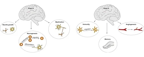
Nogo-A is one of the most potent growth inhibitors in the central nervous system. It is involved in creating new blood cells, developing new stem cells, protecting growth of cancer, and modulating the immune system. It is highly expressed after adolescent development, traumatic brain injuries, and many myelin-related diseases.
Basics

Schwab and Caroni discovered that myelin (the fatty white substance that surrounds the axon of some nerve cells) from the central nervous system (CNS) inhibits neurite outgrowth. R
Myelin from the peripheral nervous system (PNS) does the opposite. R
In the human brain, the growth of myelin is regulated by multiple systems:
oligodendrocyte-myelin glycoprotein (OMgp) R
the reticulon RTN4 (Nogo) R
semaphorins R
ephrins R
chondroitin sulphate proteoglycans R
When Nogo-A is active, it acts as a myelin-derived neurite and axon growth inhibitor. R
It suppresses growth and sprouting of neurons, thus stabilizing the wiring of the adult CNS. R
It does this by regulating axonal and neural stem cells and progenitor cells. R R
Nogo-A can also inhibit the neuronal benefits of brain derived neurotrophic factor (BDNF). R
Nogo-A And Cell Functioning
Nogo-A is involved with normal cell-functioning, along with neuropsychiatric functions. R R
It helps regulate cell death and/or growth mechanisms. R
Nogo-A can protect against hydrogen peroxide-induced cell death. R
It does this by interacting with the enzyme peroxiredoxin 2, which scavenges reactive oxygen species (ROS). R
Nogo-A is upregulated in many cells as an inhibitory factor on the growth of tumor cells, helping prevent the spread of cancer. R
Nogo-A And Injury
After injury NgR (Nogo receptors) increase. R
Nogo-A increases in the cell body of injured neurons. R
It restricts axonal regeneration after injury. R
Nogo-A In The Brain
Nogo-A is expressed in neurons throughout the brain and spinal cord (and also oligodendrocytes). R
In humans, Nogo-A has been detected in the spinal cord, in the hippocampus, in the cerebral cortex, in the cerebellum and in the brain stem. R
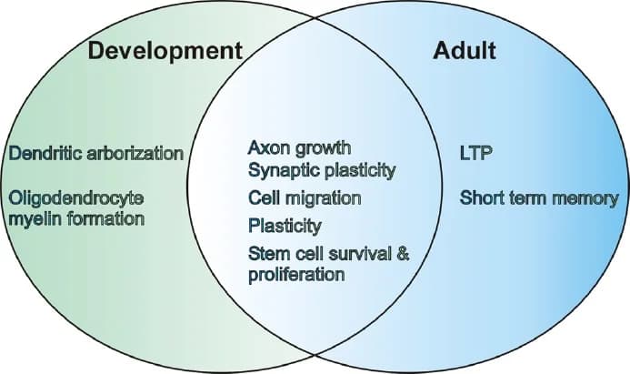
Neuronal Nogo-A is highly expressed during ages of development and down-regulated when we are adults (from birth to adolescence, it is expressed in different areas of the brain and in adulthood it is expressed more in the cerebral cortex). R R
Nogo-A expression helps the development of immature neurons before myelination. R
After myelination, Nogo-A expression is stays high in plastic CNS regions such as the hippocampus, olfactory bulb, deep cerebellar nuclei, spinal motor neurons, and dorsal root ganglia. R R
Nogo-A and Immunity
Several lymphocytes including B cells and T cells express NgR1 (Nogo-A's receptor) and further up-regulate it upon activation of the immune response. R
In the lymph nodes, T cells are activated by dendritic cellls (DCs), which express NgR1 and NgR2 during development, but are downregulated during matruation (which is inversely correlated with myelin). R
Nogo-A And Disease
Nogo-A is up-regulated in:
Benefits Of Inhibiting Nogo-A
1. May Improve Recovery After Tramautic Brain Injury

Nogo-A expression in the brain is significantly increased after stroke.R
Animals treated with anti-Nogo-A antibodies after injury have enhanced neuroplasticity and functional recovery. R R
Although, the ability of hippocampal recovery after stroke using anti-Nogo-A antibodies has been disputed. R
Anyway, inhibiting Nogo-A enhances axonal sprouting and increases dendritic complexity of neurons in the sensorimotor forelimb cortex (this area is important for skilled reaching and motor movements). R
Some studies show that treatment with Nogo-A antibodies after stroke is most effective if it is used by up to one week after injury. R R
It should be noted that inhibiting Nogo-A during stroke worsened the outcome in rodent studies (by increasing apoptosis via p53). R
In contrast, Nogo-A deficient mice that were subjected to traumatic brain injury (TBI) showed significantly worse outcomes than regular mice after TBI. R
2. May Improve Outcome of Spinal Cord Injury
Spinal cord injury (SCI) is associated with axonal disconnection. R
This leads to significant disabilities, even though there can be minimal neuronal death. R
To myelin growth regulators (MAG and OMgp) synergize with Nogo-A to restrict axonal growth after SCI. R
In mice with SCI, a Nogo-A receptor antagonist is able to increase axon regeneration. R
It also increased neuronal reorganization and behavioural improvements. R
In monkeys, Nogo-A antibodies helped improve fine motor movement recovery. R
ATI355, an anti-human Nogo-A antibody, has been shown to be safe for SCI in human clinical trials. R
3. May Improve Parkinson's Disease

Parkinson’s disease (PD) is a neurodegenerative disorder that is mainly characterized by the progressive loss of dopaminergic neurons in the substantia nigra pars compacta (SNc) with additional loss of dopamine innervation in the striatum. R
Nogo-A expression is high in the SNc in patients with PD. R
One useful strategy to replete the dopaminergic neurons in the brain is with a graft of fetal human ventral mesencepahlic dopaminergic neurons. R
For example, rats with PD were given graft transplants of dopaminergic neurons into their brain and Nogo-A inhibition made this procedure more effective (by two-fold). R
Also, antagonizing Nogo receptors significantly increased dopaminergic cell numbers. R
Tumor necrosis factor alpha (TNFa) and interleukin 6 (IL-6) are two biomarkers for inflammation during PD. R
In a model of PD, Nogo-A inhibition was able to inhibit the increase of TNFa and IL-6 (two proinflammatory cytokines) by lipopolysaccharide (LPS). R
4. Improves Multiple Sclerosis
Nogo-A activation can help identify multiple sclerosis (MS). R
Both Nogo-A and NgR1 are expressed in multiple sclerosis (MS) lesions. R
In animal models, deactivating Nogo-A expression can help ameliorate MS and promote axonal repair. R R
Using a Nogo-A antibody was able to prevent damage to the spinal cord in MS. R
5. Increases New Memory Formation

In the hippocampus, nogo-a stabilizes the architecture of the hippocampus. R
Nogo-A (along with PirB), also negatively influences long-term potentiation (LTP) in the hippocampus (via modulation of AMPA). R R R
Nogo-A influences spatial learning and memory retention by regulating the use of more efficient hippocampus-dependent strategies. R
So, inhibiting Nogo-A would theoretically allow new memories to form and overwrite old ones. R
This may be helpful for Post-Tramautic Stress Disorder, since Nogo-A expression prevents the erasure of fear memories. R
6. Protects The Eyes During Injury
Nogo-A is highly expressed in Müller glia (a type of retinal glial cell). R
It regulates inflammation and axonal growth of the optic nerve.
For example, overexpression of Nogo-A was able to help promote regeneration of retinal ganglion cells (RGCs) after optic nerve injury.R
In contrast, in multiple studies mice unable to express Nogo-A had significantly better abilities to heal their optic nerve after injury. R R R R R
For example. spatial frequency and contrast sensitivity was increased in Nogo-A deficient mice than regular mice after eye damage. R
Even inhibiting Nogo-A activity had similar results in multiple studies. R R
Also if RGCs were in an active growth state, inhibition of Nogo enhanced optic nerve regeneration even more. R
7. Promotes Angiogenesis
Nogo-A is a negative regulator of angiogensis (the growth of new blood cells). R
In mice, inhibiting Nogo-A increased blood cell formation in the brain. R
In humans, besides Nogo-A, Nogo-B regulates vascular remodeling. R
8. May Increase Healing After Peripheral Nerve Injury
Injured peripheral nerves often regenerate well, but inhibition of Nogo-A promotes their healing, especially of Schwann cells. R
9. May Prevent Hearing Loss
Nogo-A is found in sensory organs such as the inner ear. R
Nogo-A is involved in maintaining a non-regenerative state of hair cells. R
In mice, no hearing loss was observed in 10 month old Nogo-A knock-out mice as compared to wild type. R
10. May Help Amyotrophic Lateral Sclerosis
Amyotrophic lateral sclerosis (ALS) is a neurodegenerative disease characterized by motor neuron loss and muscle wasting. R
Nogo can influence the progression of ALS. R
Nogo-A expression is correlated with the severity of symptoms in ALS patients. R R R
Expression may significantly contribute to functional motor impairment. R
A Nogo-A test is able to identify ALS early in the course of the disease when diagnosis is difficult. R
In a human clinical trial, intravenous ozanezumab (anti-Nogo-A antibody) inhibited demylination of the muscle nerve fibers. R
It was also well tolerated shown to be safe. R
There are more human clinical trials showing its efficacy. R
In contrast, in an animal model, inhibiting Nogo-A promoted and worsened ALS. R
Downsides Of Inhibiting Nogo-A

in mice that had the Nogo-A gene deleted, they experienced behaviorail abnormalities resembling schizophrenia-related endophenotypes: R
deficient sensorimotor gating
disrupted latent inhibition
perseverative behavior
increased sensitivity to the locomotor stimulating effects of amphetamine
They also had altered monoaminergic transmitter levels in specific striatal and limbic structures, as well as changes in dopamine D2 receptor expression in the same brain regions. R
3. May Disrupt Circadian Rhythm
Mice lacking Nogo-A had problems with motor co-ordination and balance (via modulation of dopaminergic and motor systems). R
This was accompanied with spontaneous locomotor activity. R
Activity was increased in during the night. R
4. May Promote Cancer
Nogo-A acts as a downregulator for tumor growth. R
It is highly expressed in tumors, helping prevent their growth. R
Inhibition can theoretically allow excessive tumor growth, but I would like to see more research on this topic.
5. May Promote Alzheimer's Disease

Nogo-A/Nogo-A receptors (NgR) modulate the production of amyloid β-protein (Aβ), which is thought to be a major cause of Alzheimer's Disease (AD). R
One way it does this is through mediating neuroinflammation via modulating microglia adhesion and migration. R
The Nogo-A/NgR and the downstream Rho-ROCK pathway inhibits axon outgrowth and synapse remodeling. R
This is an obstacle to neuronal regeneration and blocking the recovery of damaged neural networks in AD. R
PirB is a novel receptor for Nogo-A that interacts with Aβ and mediates its neurotoxicity. R
S1PR2 is also a receptor for Nogo-A that activates ROCK and mediates neuronal plasticity. R
NgR also influences the metabolism of amyloid precursor protein (APP). R
NgR can bind to APP and Aβ. R
In mice, there is an increased Aβ accumulation in the hippocampal dentate gyrus and cerebral cortex of mice lacking NgR. R
Applying NgR(310)ecto-Fc (an Anti-NgR blocking protein) reduced Aβ plaque deposition in those mice. R
Also in cultures, overexpression of NgR decreases Aβ production. R
As mice aging, their ability to bing Nogo-A to Aβ decreases. R
In contrast, blocking reticulon 3 (different than reticulon 4) is beneficial for reducing Aβ. R
Also, in humans Nogo-A is over-expressed in hippocampal neurons in AD and also associated with high levels of Aβ in the hippocampus. R
Nogo is able to bind and inhibit the β-amyloid-converting enzyme 1 (BACE1), which transforms the amyloid precursor protein (APP) into aggregating β-amyloid. R
My Experience With Nogo-A
I have shown moderate amounts of Myelin-Basic Protein (MBP) antibodies and have used ginseng with good success in hopes to improve myelination via nogo-a inhibition.
How To Inhibit Nogo-A
Mechanism Of Action


Nogo-A (aka reticulon 4) belongs to the reticulon family that consists of four genes named RTN1, RTN2, RTN3 and RTN4. R
RTNs infleunce the curvature of the endoplasmic reticulum (ER) and are structural regulators for the ER. R
RTNs also interact with anti-apoptotic intracellular proteins Bcl-2 or Bcl-XL in regulating cell death. R
RTN4 encodes for three major isoforms (Nogo-A, B and C). R
Nogo-A (Nogo-66 and Nogo-A-D20) naturally binds to its receptor NgR1. R R R
NgR1 has to form a complex with LINGO-1, TROY or p75. R
p75 interacts with GPI and activates the Rho/ROCK pathway. R
Nogo-66 binding with PirB can also activate the Rho/ROCK pathway. R
Neurite growth inhibition is regulated by RhoB, Rac1, and TSPAN3 (tetraspanin-3).R R R
Nogo-A turns on RhoA, but deactivates RhoB and Rac1. R
When Nogo-A binds to Sphingolipid Receptor S1PR2, synaptic plasticity is surpressed. R
Nogo-D20 works via S1PR2. R

Nogo-A:
Nogo-A Inhibition:
More Research
Jacob Gordon
INHC, FMT-C
Board Certified Health Coach
I spent years battling unexplained chronic illness before discovering biohacking, epigenetics, and functional medicine. Now I share that research at MyBioHack to help others find their own answers.
Book a ConsultationRelated Protocols & Supplements
Deep-dive chapters and recommended supplements for this topic
Electrolyte Complex
1 scoop/day
CoQ10
200mg/day
Magnesium Glycinate
400mg at bedtime