Benefits Of Pinealon (EDR Peptide)
By Jacob Gordon, INHC, FMT-CPinealon is a synthetic tripeptide (Glu-Asp-Arg) that penetrates the cell nucleus and directly modulates gene expression in neurons, operating through a mechanism fundamentally different from receptor-mediated drugs and placing it in a category that is mechanistically unusual but biologically grounded.
In this post, we will discuss what Pinealon is, the evidence for each of its documented effects, the molecular mechanisms from DNA binding through downstream neuroprotection, the critical limitations of the evidence base, dosing, and genetics.

What Pinealon Is
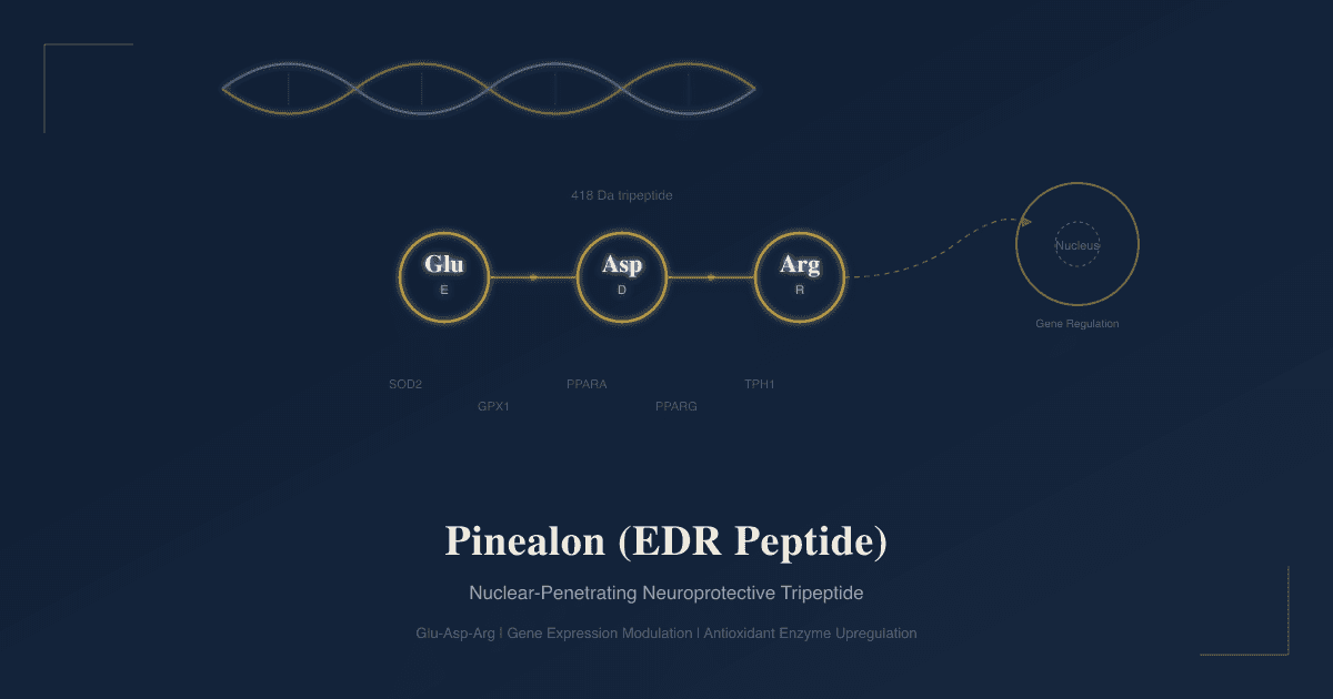
Pinealon (also called EDR peptide, sequence: Glu-Asp-Arg) is a synthetic tripeptide composed of glutamic acid (E), aspartic acid (D), and arginine (R).
Molecular weight: approximately 418 Da.
CAS: 175175-23-2.
It was isolated from Cortexin, a polypeptide neuroprotective drug derived from the cerebral cortex tissue of young cattle used clinically in Russia and Eastern Europe for stroke, traumatic brain injury, and epilepsy. R
Vladimir Khavinson's group at the St. Petersburg Institute of Bioregulation and Gerontology identified EDR as one of the shortest and most active peptide sequences within Cortexin responsible for its neuroprotective effects, then synthesized it as a standalone compound. R
Pinealon belongs to the broader class of peptide bioregulators, ultrashort (2 to 4 amino acid) signaling molecules with tissue- and gene-specific selectivity. R
Positively-charged short peptides rich in arginine have previously been established to have neuroprotective properties. Beyond excitotoxicity reduction, arginine-rich peptides diminish mitochondrial dysfunction and inhibit extracellular matrix metalloproteinase activation in neuropathology, increasing the viability of the neurovascular unit in various pathological processes. R
Naming clarification:
Despite the name, Pinealon does not come from the pineal gland. It was isolated from brain cortex extract (Cortexin), not pineal tissue. The name refers to its proposed functional relationship with neurological aging, not its tissue of origin.
The Evidence Base: What You Need To Know First
There is a MAYBE over essentially every benefit in this post, and it needs to be stated before the benefits section.
Single-source problem:
Essentially all published Pinealon research originates from one group: Khavinson's St. Petersburg Institute of Bioregulation and Gerontology and its immediate collaborators. R
No independent laboratory has replicated the core findings.
No Pinealon trials appear on ClinicalTrials.gov.
Approximately half the primary publications are in Russian only with selective English translation.
This does not mean the research is wrong. It means it has not survived the basic scientific test of independent replication. The cell-level mechanisms (antioxidant gene upregulation, MAPK/ERK timing modulation, DNA binding) are biologically plausible and consistently reported within that body of work.
Preclinical majority:
The overwhelming majority of studies use in vitro cell culture (rat cerebellar granule cells, HeLa cells, hippocampal neuron cultures) or in vivo rat models (prenatal hyperhomocysteinemia, acute hypoxia, aging, streptozotocin-induced diabetes). R R R
The human clinical data consists primarily of one reported trial in 72 patients with traumatic brain injury and one study of 59 professional truck drivers receiving peptide bioregulator correction for neurotic disorders. R R
Benefits
1. Neuroprotection Against Oxidative Stress
The brain consumes approximately 20% of total body oxygen despite comprising roughly 2% of body mass, generating substantial reactive oxygen species (ROS) as a metabolic byproduct. R
Oxidative stress is a key risk factor in Alzheimer's disease pathogenesis, with free radicals activating JNK and p38 signaling pathways that drive neuroinflammation and amyloidogenesis. R
Hydrogen peroxide is generated during the very early stages of aggregation of amyloid peptides implicated in Alzheimer's disease, making early oxidative stress a driver of amyloidogenesis rather than merely a consequence. R
Activated macrophages responding to Abeta42 aggregates produce ROS and pro-inflammatory cytokines (TNF-alpha, IL-1-beta) that activate MAPK signaling pathways and stimulate beta-secretase gene expression, creating a pro-amyloidogenic positive feedback loop. R
Pinealon suppresses ROS synthesis induced by receptor-dependent activators (ouabain, homocysteine) and non-receptor activators (hydrogen peroxide) in rat cerebellar granule cells. R
The latency period before oxidation development extends in proportion to EDR concentration: at lower doses EDR primarily reduces ROS accumulation and prevents cell death; at higher doses it shifts toward modulation of the cell cycle itself, promoting cellular proliferation. R
EDR directly neutralizes primary lipid peroxidation products and reduces hydroperoxide levels in neuronal cultures. R
EDR also reduced ROS in zymosan-activated neutrophil cultures, demonstrating antioxidant properties beyond neuronal tissue. R
Hypoxia produced a three-fold increase in initial ROS levels in neuronal cultures. Against this high-ROS background, NMDA did not cause further ROS elevation in offspring of hypoxia-exposed rats, but EDR treatment normalized both ROS levels and NMDA sensitivity. R
In hypoxia-sensitive rats, EDR elevated SOD2 (mitochondrial superoxide dismutase) and GPX1 (glutathione peroxidase 1) activity in brain tissue to levels equivalent to naturally hypoxia-resistant animals. R
2. Anti-Apoptotic Effects: Caspase-3 And p53 Suppression
Caspase-3 is the main effector caspase in neurodegeneration, driving progressive neuron death in Alzheimer's disease, cerebral ischemia, and hypoxic injury. R
Caspase-3 participates in APP processing into amyloidogenic fragments and is elevated in hippocampal axons at sites of neurofibrillary tangle and plaque formation. By cleaving Akt kinase, caspase-3 activates GSK3-beta, the primary kinase driving tau protein hyperphosphorylation. R
Pharmacological blockade of caspase-3 activation in the CNS can prevent tau phosphorylation, and drugs targeting caspase-3-dependent Akt cleavage are considered promising for preventing tau metabolism disorders in AD. R
The p53 transcription factor plays a central role in responding to DNA damage, genome integrity maintenance, and tumor suppression. R
p53 together with p63 and p73 regulates the cell cycle, apoptosis, differentiation, and cell aging. Disruption of p53 activity leads to NDDs, cancer, and metabolic syndrome. R
Overexpression of p53 has been confirmed in cortical neurons of frontal and temporal lobes, glial cells, and hippocampus in both familial and sporadic AD, where it correlates with Abeta42 accumulation. R
EDR peptide reduced caspase-3 expression in brain tissue and improved learning performance in the Morris water maze in both young and old rats under acute hypoxic hypoxia. R
In old rats, acute hypoxia increased caspase-3 activity in brain neurons; EDR administration prevented this activation, maintaining neuronal viability. R
3. MAPK/ERK Signaling Pathway Modulation
Mitogen-activated protein kinases (MAPKs) are evolutionarily conserved signaling molecules converting extracellular signals into intracellular responses. The four main mammalian MAPKs are ERK1/2, JNK, p38, and ERK5. R
In cerebellar granule cells exposed to homocysteine (a MAPK activator and pro-oxidant), ERK1/2 activated within 2.5 minutes in controls. In the presence of EDR peptide, ERK1/2 activation was delayed to 20 minutes, a 17.5-minute lag-phase extension. R
This temporal shift changes the onset of cell cycle phases: lower EDR concentrations limit ROS accumulation and prevent cell death; higher concentrations modulate the cell cycle itself. R
4. SOD2 And GPX1 Antioxidant Enzyme Upregulation
SOD2 is located in mitochondria and is the only ROS-regulated isoform of superoxide dismutase. Mice lacking SOD2 develop neuropathology through neuronal apoptosis caused by mitochondrial oxidative stress. R
EDR has binding sites in promoter regions of both SOD2 and GPX1 genes confirmed by molecular modeling, and functionally elevates SOD2 and GPX1 activity in hypoxia-sensitive rat brains to levels equivalent to hypoxia-resistant animals. R
5. PPARA And PPARG Transcription Factor Activation
PPARA and PPARG are nuclear ligand-activated receptors with anti-inflammatory and anti-amyloidogenic roles in the brain. R
PPARA activation induces proteolysis of APP by alpha-secretase in hippocampal neurons by inducing ADAM10 gene transcription, shifting APP processing away from the amyloidogenic beta-secretase/gamma-secretase pathway. R
EDR peptide increased expression of PPARA and PPARG genes in humans under physical exercise-induced stress conditions. R
Molecular modeling identified three EDR binding sites in the PPARA gene promoter and five in the PPARG gene promoter. R
6. Serotonin Synthesis Upregulation Via TPH1
In AD, serotonin concentration decreases in the anterior cortex, hippocampus, amygdala, and striatum. Dysfunction of serotonergic signaling leads to amyloidogenesis, tau hyperphosphorylation, and neurofibrillary tangle formation. R
EDR peptide and the related peptide Lys-Glu-Asp both stimulate serotonin expression in aging cultures of brain cortex cells. R
Peptide regulation of the TPH1 gene was demonstrated by molecular docking, with the CCTGCC nucleotide sequence in the TPH1 gene found to be complementary to EDR. R
7. Dendritic Spine Preservation In Alzheimer's And Huntington's Disease Models
Mushroom-shaped dendritic spines form active synapses and are representative of a highly integrated neural network that serves as the anatomical substrate for learning and memory. R
In hippocampal neuron cultures derived from 5xFAD AD mice, EDR peptide prevented the loss of mushroom-shaped dendritic spines that occurs in the disease model. R
In a Huntington's disease mouse model, EDR peptide restored the morphology of spines in striatal neurons. R
8. Prenatal And Developmental Neuroprotection
In a rat model of prenatal hyperhomocysteinemia induced by dietary methionine loading during pregnancy, Pinealon administration (10 mcg/kg for 5 days) to pregnant rats produced offspring with significantly improved spatial orientation, improved Morris water maze learning, decreased ROS accumulation, and decreased necrotic cell counts in cerebellar granule cell populations. R
The protective effect was attributed to reduction of the neurotoxic influence of homocysteine on the developing brain rather than by lowering homocysteine blood levels, confirming that Pinealon acts at the neural tissue level. R
9. Diabetes-Related Cognitive Impairment And NMDA Receptor Protection
Pinealon at doses of 50, 100, and 200 ng/kg showed dose-dependent effects on maintenance of previously acquired spatial learning skills in Morris water maze following streptozotocin-induced diabetes. The most positive effect was at 100 ng/kg. R
Pinealon also modulated the subunit composition of NMDA receptors in the hippocampus of diabetic rats. R
10. Anti-Hypoxic Properties And Short Peptide Neuroprotection
Investigation of anti-hypoxic properties of short peptides (including EDR) demonstrated protective effects on brain neurons exposed to hypoxic conditions in vivo. R
The excitotoxicity-protective effect is directly relevant to clinical hypoxia conditions including stroke, traumatic brain injury, and ischemic episodes where NMDA overactivation drives secondary neuronal death following the primary insult. R
11. Occupational Stress: Truck Driver Study
A study of 59 professional long-haul truck drivers treated with peptide bioregulator correction (including Pinealon) for neurotic disorders documented improvements in neurological stress markers, adaptive potential, resistance to occupational stress, and reduction in neurotic disorder risk. R
12. Traumatic Brain Injury Recovery: Human Clinical Data
Oral administration of Pinealon as adjunct to standard therapy in 72 patients with traumatic brain injury consequences and cerebrasthenia produced: R
- Improved memory
- Reduced duration and intensity of headaches
- Emotional balance
- Enhanced performance efficacy
- Decreased number of errors on the correction test
- Significant increase in the alpha-index in bioelectric brain activity (EEG)
The full trial methodology (randomization, blinding status, control design) is not well-documented in accessible English-language publications, which limits interpretation of this finding.
13. Calmodulin Modulation
Calmodulin is a calcium-sensing protein functioning as a multifunctional intracellular messenger that regulates over 300 cellular proteins. R
EDR peptide affects calmodulin synthesis in neurons, representing an additional molecular target of the peptide's gene-regulatory activity. R
Dosage And Administration
No large, well-controlled pharmacokinetic dose-finding study has been conducted for Pinealon in humans. The following derives from published clinical reports and the Khavinson research group's documented protocols.
Oral (reported in human TBI trial):
0.2 mg twice daily for 20 to 30 days. R
Sublingual:
Holding solution under the tongue for 30 to 60 seconds improves mucosal absorption, bypassing first-pass gastrointestinal peptide degradation. No formal sublingual bioavailability study for Pinealon specifically has been published.
Subcutaneous injection:
100 to 300 mcg daily. This route provides the most direct delivery and avoids gastrointestinal peptide degradation.
Animal model reference doses:
The prenatal hyperhomocysteinemia rat study used 10 mcg/kg daily for 5 days. R
The diabetes/NMDA rat study used 50, 100, and 200 ng/kg for 5 days, with 100 ng/kg showing best outcomes. R
Cycling:
Following the Khavinson peptide bioregulator model, most practitioners use 10 to 20 consecutive days every 3 to 6 months rather than continuous daily use.
Safety:
No serious adverse effects have been reported in the published literature at clinical doses. EDR is not immunogenic due to its small size and absence of species-specificity. The arginine content at these doses is negligible in terms of dietary arginine load.
Mechanisms Of Action
Simple:
- Pinealon's small size (418 Da) and arginine content allow it to cross both cell membranes and the nuclear membrane, reaching the cell nucleus without requiring a surface receptor R
- Once in the nucleus it binds to specific DNA promoter sequences (d(CCTGCC)2 and d(CCAGC)2), upregulating transcription of SOD2, GPX1, PPARA, PPARG, and TPH1, the genes governing mitochondrial antioxidant defense, neuroinflammation control, and serotonin synthesis R R
- It delays MAPK/ERK1/2 activation in neurons under oxidative stress from 2.5 minutes to 20 minutes, changing cell cycle phase timing in a way that reduces ROS accumulation and prevents apoptosis at lower doses R
- It suppresses caspase-3 and p53 expression, directly reducing the rate of programmed neuronal death under hypoxic and oxidative conditions and indirectly preventing tau hyperphosphorylation via the Akt-GSK3-beta axis R R
- It preserves mushroom-shaped dendritic spines in Alzheimer's and Huntington's disease models, maintaining the anatomical substrate for synaptic plasticity and memory R R
- It epigenetically upregulates TPH1 transcription, increasing serotonin synthesis in aging brain cortex cells and counteracting the serotonin depletion characteristic of neurodegeneration R
- It activates PPARA and PPARG, which shift APP processing toward the non-amyloidogenic alpha-secretase pathway via ADAM10 induction and suppress neuroinflammatory cytokine production R
Advanced:
Nuclear membrane penetration and DNA interaction:
Fluorescence-labeled EDR peptide studies in HeLa cells confirmed translocation across both cellular and nuclear membranes, establishing that the peptide reaches the cell nucleus rather than acting at a surface receptor. R
In vitro studies showed EDR interacts with double-stranded DNA, exerting a destabilizing effect on its secondary structure and a compacting effect on its molecular coil volume, consistent with intercalation-adjacent binding mechanics. R
Molecular modeling identified two primary binding motifs (d(CCTGCC)2 and d(CCAGC)2) appearing in promoter regions of PPARA (three binding sites), PPARG (five binding sites), SOD2, GPX1, and TPH1 genes. R
MAPK/ERK cascade in AD:
EDR's 17.5-minute lag-phase delay of ERK1/2 activation under homocysteine exposure is not suppression but a temporal shift: it preserves the eventual proliferative and differentiation functions of ERK1/2 while preventing the early pro-apoptotic ROS-driven phase from triggering irreversible cell death. R
PPARA-ADAM10-alpha secretase axis:
PPARA activation induces ADAM10 gene transcription, increasing alpha-secretase availability and shifting APP processing toward the non-amyloidogenic pathway. By upregulating PPARA expression, EDR provides an indirect but mechanistically coherent route to reduced Abeta42 production. R
Caspase-3 and GSK3-beta-tau axis:
By cleaving Akt, caspase-3 activates GSK3-beta, which phosphorylates tau protein to produce neurofibrillary tangles. EDR's suppression of caspase-3 is relevant not only to preventing cell death but to preventing two AD hallmarks: amyloidogenic fragment accumulation and neurofibrillary tangle formation. R
Genetics
SOD2 Val16Ala (rs4880):
Val/Val genotype results in less efficient mitochondrial SOD2 import, lower mitochondrial antioxidant capacity, and higher baseline mitochondrial oxidative stress. Given that EDR has a confirmed SOD2 promoter binding site and functionally elevates SOD2 activity, Val/Val carriers (lowest baseline mitochondrial SOD2) are theoretically most sensitive to EDR's antioxidant upregulation. R
GPX1 Pro198Leu (rs1050450):
GPX1 Leu/Leu carriers have reduced GPX1 enzyme activity, higher susceptibility to hydrogen peroxide accumulation, and greater lipid peroxidation vulnerability. EDR has a GPX1 promoter binding site and functionally elevates GPX1 activity, making Leu/Leu carriers the logical population most responsive to EDR's antioxidant-upregulating effects. R
PPARA and PPARG variants:
PPARA rs1800206 (Leu162Val) reduces PPARA transcriptional activity. PPARG Pro12Ala (rs1801282, Ala allele) is associated with reduced PPARG activity. EDR's documented upregulation of both PPARA and PPARG is of potential particular relevance in individuals with constitutively lower expression of these transcription factors. R
MTHFR C677T (rs1801133):
MTHFR TT homozygotes have significantly elevated homocysteine levels. The most robust Pinealon neuroprotective finding was in the prenatal hyperhomocysteinemia model, and EDR suppresses homocysteine-induced ERK1/2 activation and ROS production in neurons. MTHFR TT carriers with chronically elevated homocysteine may be the population most directly analogous to the rat model in which EDR showed its most robust neuroprotective effects. R R
APOE4, APP, PSEN1, PSEN2:
For AD-specific effects, APOE4 carriers and familial AD mutation carriers have the most dysregulated versions of the pathways EDR targets: elevated beta-secretase activity (amenable to PPARA-ADAM10 correction), increased oxidative stress (amenable to SOD2/GPX1 upregulation), and accelerated caspase-3-driven tau pathology (amenable to direct caspase-3 suppression). No human trial has been conducted.
TPH1 variants (rs1800532, rs1799913):
Low-activity TPH1 variants associated with reduced serotonin synthesis capacity compound the serotonin depletion seen in aging and neurodegeneration. EDR's TPH1 promoter binding and serotonin upregulation are most relevant for individuals whose baseline TPH1 transcription is already reduced by genetic variation. R
More Research
- Independent replication is the defining gap. The mechanistic case for Pinealon across its documented targets (SOD2/GPX1 upregulation, MAPK timing modulation, caspase-3 suppression, PPARA/PPARG activation, TPH1-serotonin upregulation, dendritic spine preservation) is internally consistent and biologically plausible. R No independent laboratory has confirmed any finding. The most immediately testable priority is SOD2/GPX1 promoter binding and functional enzyme upregulation, which could be confirmed with routine cell biology methods without animal models or clinical trials.
- 5xFAD dendritic spine finding warrants independent follow-up. EDR's prevention of mushroom-spine loss in hippocampal neurons from 5xFAD mice R and restoration of spine morphology in striatal neurons in the Huntington's model R are the most clinically translatable findings in the literature. Independent replication using rigorous spine morphology analysis would substantially change the evidence picture.
- Diabetic cognitive impairment is an underexplored application. The streptozotocin diabetes study showing Pinealon's dose-dependent preservation of spatial memory and hippocampal NMDA receptor subunit normalization R is mechanistically relevant to the large and growing population with type 2 diabetes-associated cognitive decline. No human trial has examined Pinealon in this context.
- Post-viral neurological syndromes. The mechanisms EDR modulates (NF-kB, PPARA/PPARG, caspase-3, MAPK-JNK/p38, NMDA excitotoxicity, neuronal oxidative stress) overlap substantially with pathological features of post-viral neurological syndromes including long COVID-associated brain fog and neuroinflammation. No research has examined Pinealon in this context. The mechanistic overlap is present and represents an unaddressed but tractable research question.
Jacob Gordon
INHC, FMT-C
Board Certified Health Coach
I spent years battling unexplained chronic illness before discovering biohacking, epigenetics, and functional medicine. Now I share that research at MyBioHack to help others find their own answers.
Book a ConsultationRelated Protocols & Supplements
Deep-dive chapters and recommended supplements for this topic
Lion's Mane
1000mg/day
Omega-3 (DHA)
2g/day
Phosphatidylserine
100mg 3x/day